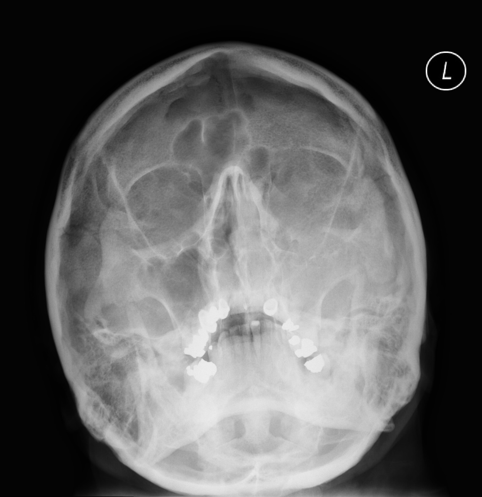
Orbital Roof Fracture Radiology

Pin On Mr Radiology

Orbital Roof Blow In Fracture Radiology Case Radiopaedia Org

Blowout Orbital Fracture Radiology Medical Imaging Radiography

Osteoma Of Orbital Roof Radiology Case Radiopaedia Org Radiology Brain Images Mri Brain

Superior Orbital Fracture Radiology Case Radiopaedia Org

Pathological Clavicle Fracture Radiology Case Radiopaedia Org Radiology Pathology Clavicle
Isolated orbital roof fractures in adults are uncommon comprising 12 19 of all orbital wall fractures.
Orbital roof fracture radiology.

Osteoma Of Orbital Roof Radiology Case Radiopaedia Org Radiology Radiology Imaging Diagnostic Imaging

Superior Orbital Roof Blowout Fracture With Intact Orbital Rim Radiology Case Radiopaedia Org

Radial Head Fracture Radiology Case Radiopaedia Org Radiology Fractures Medical School Stuff

Pin On Radiology Spotters

Facial Bone X Ray Lateral View Www Anatomynote Com Facial Bones Radiology Radiology Humor

Fibrous Dysplasia Frontal A And Lateral B Plain Radiographs Show A Well Defined Thickened And Sclerotic Appearance Of The R Patient Radiology Radiographer

Multiple Myeloma Is The Most Common Primary Malignant Bone Neoplasm In Adults And Results In A Wide Range Of Ra Radiology Multiple Myeloma Pediatric Radiology

Derrick Rose Orbital Fracture Surgery Derrick Rose Plastic And Reconstructive Surgery Head And Neck

Sacrococcygeal Giant Cell Tumour Radiology Case Radiopaedia Org Tumor Radiology Cell

Blow Out Fracture The Most Common Portion Of The Orbit To Sustain A Fracture Is The Weak Floor And This Injury If Occurring In Isolation May Result In A Blow

Maxillofacial Injuries Radiology Key

Pin On Radiologie

Pin En Radiology Interesting Cases And Spotters

Vocal Cord Paralysis Radiology Reference Article Radiopaedia Org In 2020 Thyroid Surgery Paralysis Nerve Palsy

Chest Ct Scan In A Patient Who Was Born With Only One Lung Arrowhead Normal Lung Arrow Heart Shifted To Where O Diagnostic Imaging Radiology Ct Scan

Colles Fracture Posterior Displacement

Burst Fracture Of Lumbar Spine Sagittal Reconstruction Of Ct Of The Lumbar Spine Demonstrates A Comminuted Vertical Burst Fracture Through The Body Of L1 Whit

Mount Fuji Sign Is Seen On Cross Sectional Imaging And Implies Tension Pneumocephalus Is Present The Sign Refers To The Pre Radiology Sinusitis Brain Images
1

Pin On Surgery

Sclerotic Bone Leison Note Sclerotic Bone Leison In Tibia In Young Age Osteoid Osteoma Note Malignancy In Old Age Radiology Tumor Oral Pathology

Subdural Acute On Chronic Radiology Case Radiopaedia Org Radiology Medical Imaging Brain Images

Infantile Head Trauma With Orbital Roof Fracture Radiology Case Radiopaedia Org

Pin On Radiology Study
Source : pinterest.com
